Beyond cancer: biomarkers open a new era for PET
Time zone : Europe/Brussels
Month : 2026-09
Cognitive impairment, breathlessness or persistent inflammation: similar symptoms may reflect different biological mechanisms. Identifying them becomes essential as treatments become more specialised. Positron emission tomography — PET — contributes to this approach beyond oncology. By locating a target or measuring a function, it connects biology with the affected organ. New radiotracers are expanding this approach, although their levels of validation still vary considerably.
From diagnosing a disease to understanding its mechanism
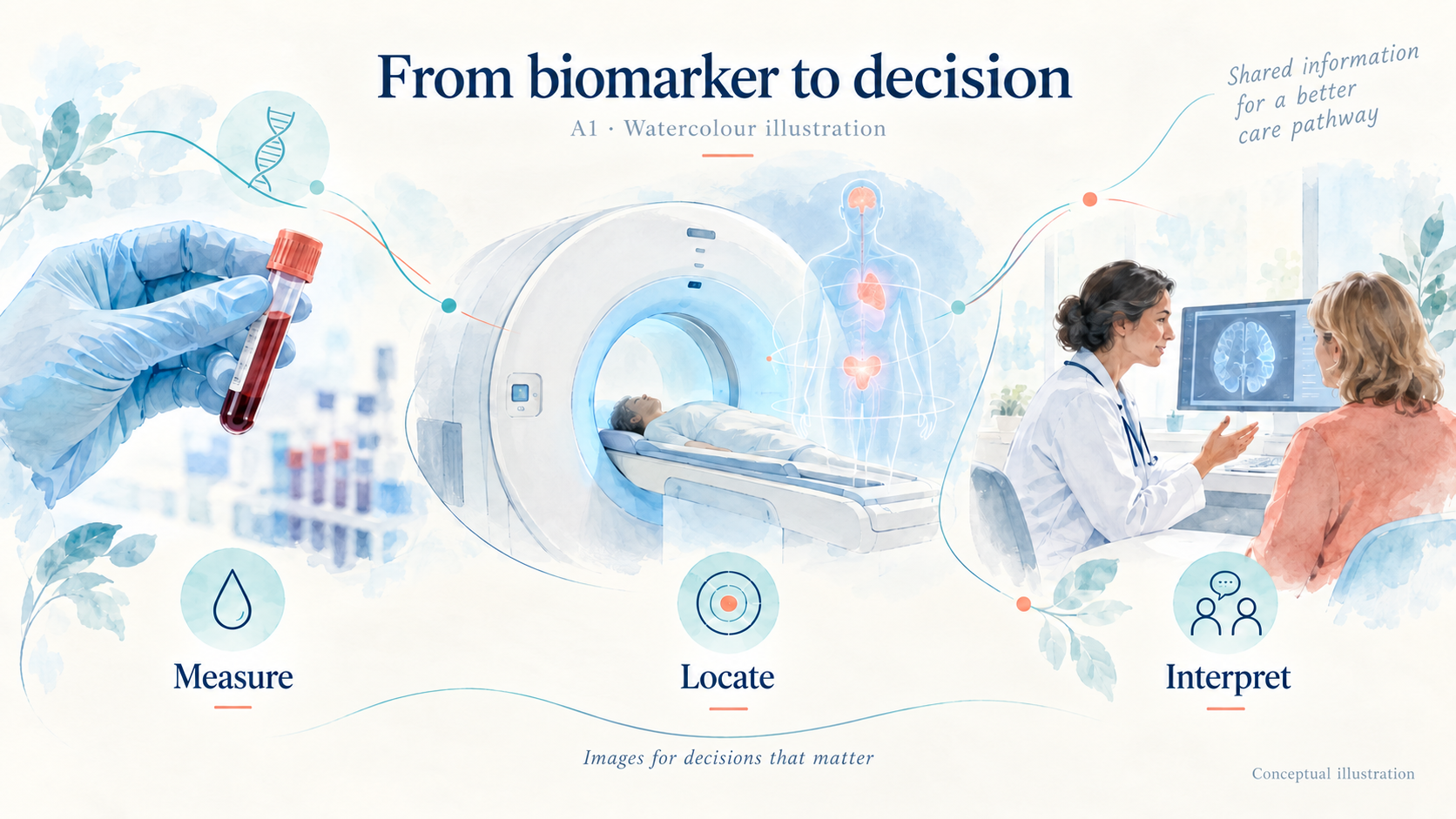
Medicine has long used biomarkers: blood glucose, troponin and inflammatory proteins. Molecular imaging can investigate protein accumulation, cellular activity or changes in blood flow within tissues. It thus complements clinical and laboratory information.
This development calls for a distinction between symptoms, lesions and mechanisms. An anatomical abnormality describes what is visible in an organ; a biomarker may provide information about a process contributing to that abnormality. These two perspectives must remain connected, since neither necessarily explains the entire clinical picture on its own.
A biomarker may contribute to diagnosis, prognosis, treatment selection or monitoring. These functions are not interchangeable: the presence of a target does not guarantee that a drug will be effective; a change in signal does not, on its own, demonstrate clinical benefit.
The radiotracer is the molecule administered; the imaging biomarker is the information measured from its distribution. This distinction requires us to specify what the signal represents and which decision it can inform.
Blood biomarkers and PET: two levels of information
Blood sampling makes repeated measurements easier. PET provides spatial information: the distribution and extent of an abnormality and heterogeneity between tissues. Combining them should address a clinical question, without routinely multiplying investigations.
The Alzheimer’s Association’s 2025 guidelines concern patients with objective cognitive impairment assessed in specialist care. They suggest a blood-based triage test if its sensitivity is at least 90% and its specificity at least 75%. A test achieving at least 90% for both parameters may replace amyloid PET or cerebrospinal fluid biomarkers. These recommendations are conditional, with low certainty of evidence; they do not extend to screening asymptomatic individuals. Blood-based biomarker guidelines, 2025. Sources: https://alz-journals.onlinelibrary.wiley.com/doi/10.1002/alz.70535
The choice therefore depends on the performance of the actual test used, the pre-test probability of disease and the remaining uncertainty. A laboratory result does not remove the need for clinical assessment.
Alzheimer’s disease: moving towards a biological understanding
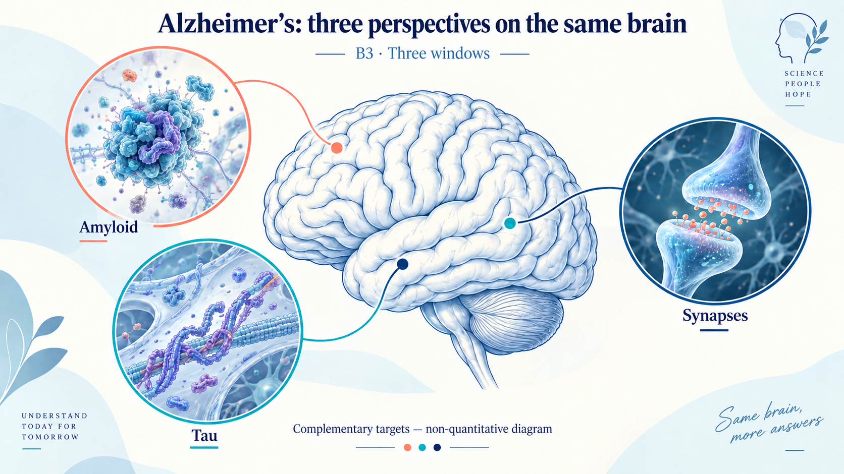
Three amyloid tracers have European marketing authorisation: [¹⁸F]florbetapir, Amyvid, since 14 January 2013; [¹⁸F]florbetaben, Neuraceq, since 20 February 2014; and [¹⁸F]flutemetamol, Vizamyl, since 22 August 2014. They assess the density of β-amyloid neuritic plaques in adults with cognitive impairment being evaluated for Alzheimer’s disease or other causes. The examination complements clinical assessment; positive uptake alone does not establish the diagnosis. EMA: Amyvid, Neuraceq, Vizamyl. Sources: https://www.ema.europa.eu/en/medicines/human/EPAR/amyvid ; https://www.ema.europa.eu/en/documents/product-information/neuraceq-epar-product-information_en.pdf ; https://www.ema.europa.eu/en/medicines/human/EPAR/vizamyl
The appropriate use criteria published in January 2025 define the role of amyloid and tau PET across diagnostic and therapeutic scenarios. They do not justify automatic referral for every memory complaint. Alzheimer’s Association/SNMMI criteria, 2025. Sources: https://doi.org/10.1002/alz.14338
For tau, [¹⁸F]flortaucipir, Tauvid, has been authorised in the United States since 28 May 2020 and in the European Union since 22 August 2024. The European indication concerns the neocortical distribution of tau neurofibrillary aggregates in adults with cognitive impairment being evaluated for Alzheimer’s disease, as an adjunct to other assessments. FDA and EMA: Tauvid. Sources: https://www.accessdata.fda.gov/drugsatfda_docs/appletter/2020/212123Orig1s000Ltr.pdf ; https://www.ema.europa.eu/en/medicines/human/EPAR/tauvid
Beyond Alzheimer’s: observing synapses and pathological proteins
[¹⁸F]SynVesT-1 targets the synaptic vesicle protein SV2A. Its first human evaluation, published online in 2020 and in the 2021 volume, established the feasibility of this imaging approach. The signal provides information about a synaptic target; it does not directly count neurons. Its use remains primarily in research. Naganawa and colleagues. Sources: https://doi.org/10.2967/jnumed.120.249144
For alpha-synuclein, the study by Smith and colleagues, published in 2023, showed [¹⁸F]ACI-12589 uptake characteristic of multiple system atrophy, but limited uptake in sporadic Parkinson’s disease. A shared protein target therefore does not guarantee identical performance across diseases. Nature Communications, 2023. Sources: https://www.nature.com/articles/s41467-023-42305-3
These approaches could clarify coexisting mechanisms in an individual patient. Their clinical utility still requires validation beyond simply visualising a signal.
In cardiology, a biomarker may be a measure of function
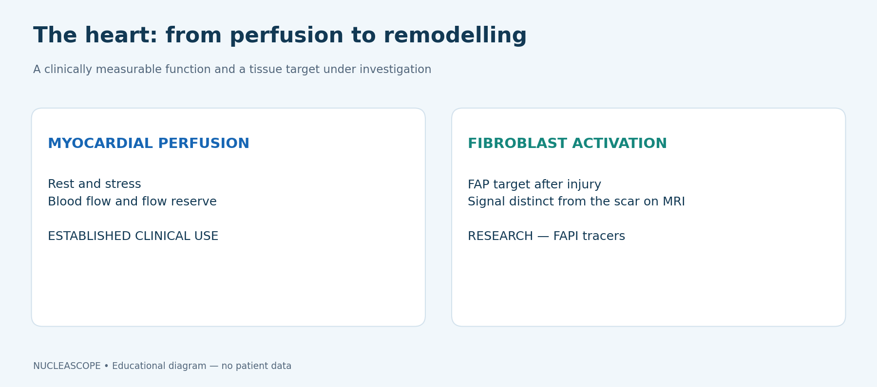
Myocardial blood flow and myocardial flow reserve under stress are functional biomarkers. Perfusion PET can quantify them beyond visual comparison between territories. The 2024 ESC guidelines position this approach for characterising ischaemia, assessing risk and measuring myocardial blood flow, according to clinical likelihood and available resources. ESC guidelines, 2024. Sources: https://doi.org/10.1093/eurheartj/ehae177
Reduced reserve does not, however, identify a single cause: epicardial coronary disease, microvascular dysfunction and haemodynamic conditions must be considered together. The value of the measurement lies in this interpretation, integrated with anatomy and symptoms.
This makes the question addressed to the nuclear medicine physician particularly important. Looking for ischaemia and understanding the origin of functional limitation are not exactly the same process; a quantitative result must be interpreted in the context that prompted the examination.
[¹⁸F]flurpiridaz, Flyrcado, was approved by the FDA on 27 September 2024 for myocardial perfusion PET at rest or under pharmacological or exercise stress, to evaluate ischaemia and infarction in adults with known or suspected coronary artery disease. It joins a field that already uses, among others, [⁸²Rb]rubidium and [¹³N]ammonia. FDA: indication and approval letter. Sources: https://www.fda.gov/drugs/news-events-human-drugs/fda-approves-imaging-drug-evaluation-myocardial-ischemia-and-infarction ; https://www.accessdata.fda.gov/drugsatfda_docs/appletter/2024/215168Orig1s000Ltr.pdf
Cardiac amyloidosis: choosing the right technique, not necessarily the newest
The ESC’s 2021 position statement recalls the role of bone-tracer scintigraphy, with tomographic confirmation of myocardial uptake. Grade 2 or 3 uptake can contribute to a non-invasive diagnosis of transthyretin amyloidosis in a compatible context, after a monoclonal abnormality has been excluded by appropriate tests. A monoclonal abnormality requires a different diagnostic approach, particularly to distinguish AL from ATTR. This is not PET. ESC position statement on cardiac amyloidosis, 2021. Sources: https://doi.org/10.1093/eurheartj/ehab072
A technique’s novelty is therefore not its main measure of value. It must add useful information to an already validated diagnostic pathway.
Inflammation: FDG retains a central role
[¹⁸F]fluorodeoxyglucose, or [¹⁸F]FDG, retains a major clinical role in infection and inflammation. Version 2.0 of the EANM/SNMMI guidelines was published online in 2024 and in the 2025 volume. The signal reflects metabolic activity, without identifying an infection or its cause on its own. EANM/SNMMI guidelines. Sources: https://doi.org/10.1007/s00259-024-06915-3
Blood glucose, treatments, preparation and acquisition timing determine its interpretation. Uptake may be inflammatory, infectious, tumour-related or physiological.
Cardiac sarcoidosis illustrates this dependence on protocol: insufficient suppression of physiological myocardial glucose consumption can obscure interpretation. PET looks for active inflammation; interpretation is integrated with perfusion findings, MRI and the clinical context. Diffuse uptake should not automatically be regarded as pathological. EANM/SNMMI, sarcoidosis section. Sources: https://doi.org/10.1007/s00259-024-06915-3
For large-vessel vasculitis, the 2023 EULAR update, published in the 2024 volume, recommends ultrasound as the first-line investigation in suspected giant cell arteritis, including the axillary arteries. PET or MRI are alternatives. If relapse is suspected, imaging may help when inflammatory markers are unreliable; it is not routine follow-up for every patient. EULAR recommendations. Sources: https://doi.org/10.1136/ard-2023-224543
Fibrosis: seeing the activity that builds the scar
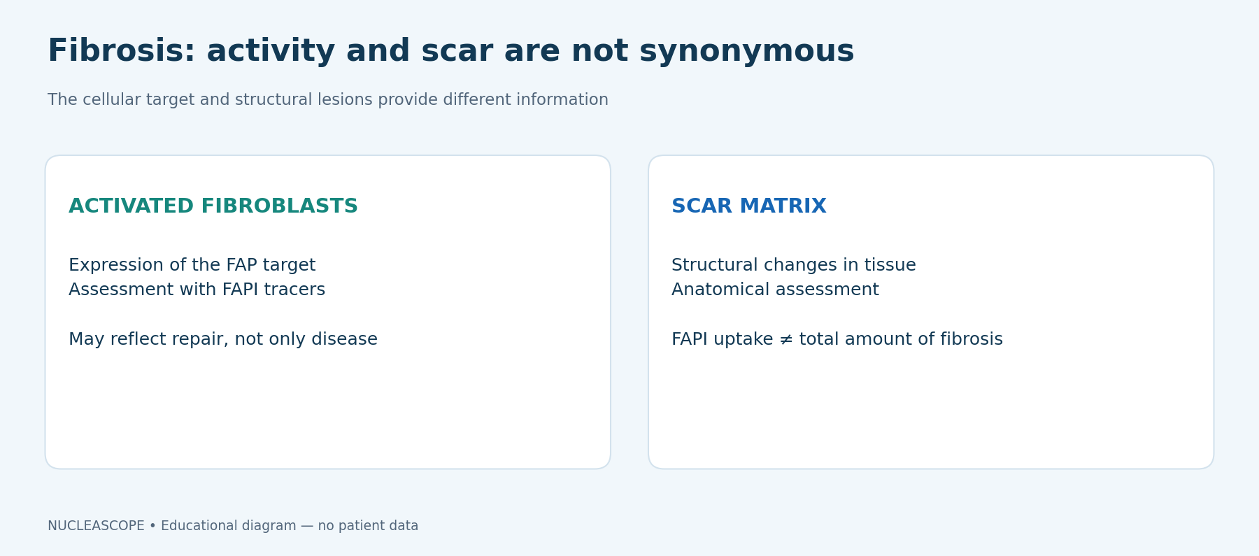
Established fibrosis and an active fibrosing process are not the same phenomenon. Fibroblast activation protein inhibitors, or FAPI, investigate a target associated with activated fibroblasts. Their non-oncological development focuses, among other areas, on the lung and cardiac remodelling.
In an exploratory study published in 2024, four patients with advanced interstitial lung disease underwent [⁶⁸Ga]FAPI-46 PET before lung transplantation. Analysis of tissue samples from three patients showed a correlation between uptake and immunohistochemical FAP expression. This small sample supports initial biological validation, without demonstrating that imaging-guided treatment improves prognosis. Hotta and colleagues, 2024. Sources: https://doi.org/10.2967/jnumed.124.268351
The study by Barton and colleagues, published in February 2025, combined [⁶⁸Ga]FAPI-46 and MRI after myocardial infarction. It investigates the distribution and evolution of fibroblast activation, distinct from the scar visualised by late gadolinium enhancement. This is research into remodelling, not a routine indication for PET after infarction. JACC, 2025. Sources: https://doi.org/10.1016/j.jacc.2024.10.103
FAPI uptake does not measure all fibrosis. Tissue repair and benign processes can also activate fibroblasts. Interpretation therefore requires anatomical and clinical correlation; an area of uptake does not automatically mean progressive disease. Pitfalls and non-tumour uptake, 2022. Sources: https://pmc.ncbi.nlm.nih.gov/articles/PMC9157730/
The research question is now whether this information usefully complements structural and functional measurements, and in which situations. For monitoring, the meaning of a change in uptake must also be established: a change in the target, a treatment effect or a measurement difference. This step separates a marker’s biological validation from its validation as a decision-making tool.
Infection: getting closer to the microorganism
Even before newer tracers, endocarditis is already an important decision-making application of [¹⁸F]FDG PET. The 2023 ESC guidelines include it in multimodality imaging for possible prosthetic valve endocarditis. It may also be considered for possible infection related to a cardiac implantable electronic device. The examination complements echocardiography and microbiology; it does not replace them. ESC endocarditis guidelines, 2023. Sources: https://www.escardio.org/guidelines/clinical-practice-guidelines/all-esc-practice-guidelines/endocarditis/
Extracardiac assessment can look for distant infectious foci. Interpretation must account for postoperative changes and context: not every area of uptake around implanted material represents infection. The strategy requires multidisciplinary discussion, especially when initial examinations remain equivocal. EANM/SNMMI, version 2.0. Sources: https://doi.org/10.1007/s00259-024-06915-3
[¹⁸F]fluorodeoxysorbitol, or [¹⁸F]FDS, explores a different pathway: specific microbial mechanisms rather than the host response alone. A prospective study published in 2021 evaluated this approach in Enterobacterales infections and control situations. These findings remain part of clinical development. Ordonez and colleagues, 2021. Sources: https://doi.org/10.1126/scitranslmed.abe9805
In 2025, Ruiz-Gonzalez and colleagues combined preclinical work with an initial evaluation in nine patients with suspected invasive mould infection, including four confirmed infections. This proof of concept does not make FDS a universal detector of bacteria or fungi. Microbiological identification and susceptibility to treatment remain essential. Nature Communications, 2025. Sources: https://doi.org/10.1038/s41467-025-61700-6
The challenge is to define when additional non-invasive information would genuinely be useful: a focus that is difficult to sample, multiple lesions or discordant investigations. Initial studies do not allow performance to be extrapolated to all microorganisms, all infection sites or all immunocompromised patients.
From Europe to the rest of the world: development at different speeds
Scientific validation, marketing authorisation, availability and reimbursement are separate steps. A publication does not constitute authorisation; authorisation does not guarantee supply or funding in every country.
It is also necessary to reason by indication. Authorisation of a brain tracer does not automatically validate its use in another organ. Research access at an expert centre does not imply routine use.
For a referring clinician, a request should therefore specify the clinical question and the tracer being considered. For the patient, useful information also includes what the examination can actually establish and which uncertainties will remain afterwards.
The overview summarises the examples presented; it is not an exhaustive worldwide inventory of authorisations.
Maturity at a glance Cerebral amyloid — [¹⁸F]florbetapir, [¹⁸F]florbetaben, [¹⁸F]flutemetamol — Marketing authorisation in the European Union, for the defined indications. Cerebral tau — [¹⁸F]flortaucipir — Tauvid: authorised in the United States in 2020 and in the European Union in 2024, for the defined indications. Myocardial perfusion — Blood flow and flow reserve; [¹⁸F]flurpiridaz — Established clinical use of perfusion PET; Flyrcado authorised in the United States in 2024. Infection and inflammation — [¹⁸F]FDG — Clinical uses governed by guidelines; non-specific signal, protocol is crucial. Synapses and alpha-synuclein — [¹⁸F]SynVesT-1, [¹⁸F]ACI-12589 — Research; performance depends on the disease and target. Fibroblast activation — FAPI, including [⁶⁸Ga]FAPI-46 — Clinical research in the non-oncological indications presented. Certain microbial infections — [¹⁸F]FDS — Clinical development; not a universal infection test.
What will make a difference for patients
A biomarker’s value lies in the uncertainty it can resolve. Before guiding treatment, it must be reproducible, validated in the population concerned and compared with available examinations.
For PET beyond oncology, the aim is to translate localised biological information into a useful decision. Discussion between specialties remains essential to interpret the signal, its limitations and its practical consequences for the patient.