The rise of PET imaging: twenty years of transformation in oncology
Month : 2026-05
From a last-resort modality to decision-driving imaging, PET has profoundly reshaped diagnostic, prognostic and theranostic standards in cancer care.
Few medical techniques will have experienced, in the space of two decades, a trajectory as rapid as positron emission tomography (PET). Born in the research laboratories of the 1970s–1980s, long confined to university centers due to lack of wide access to radiotracers and hybrid equipment, it has gradually established itself as a pillar of modern oncology.
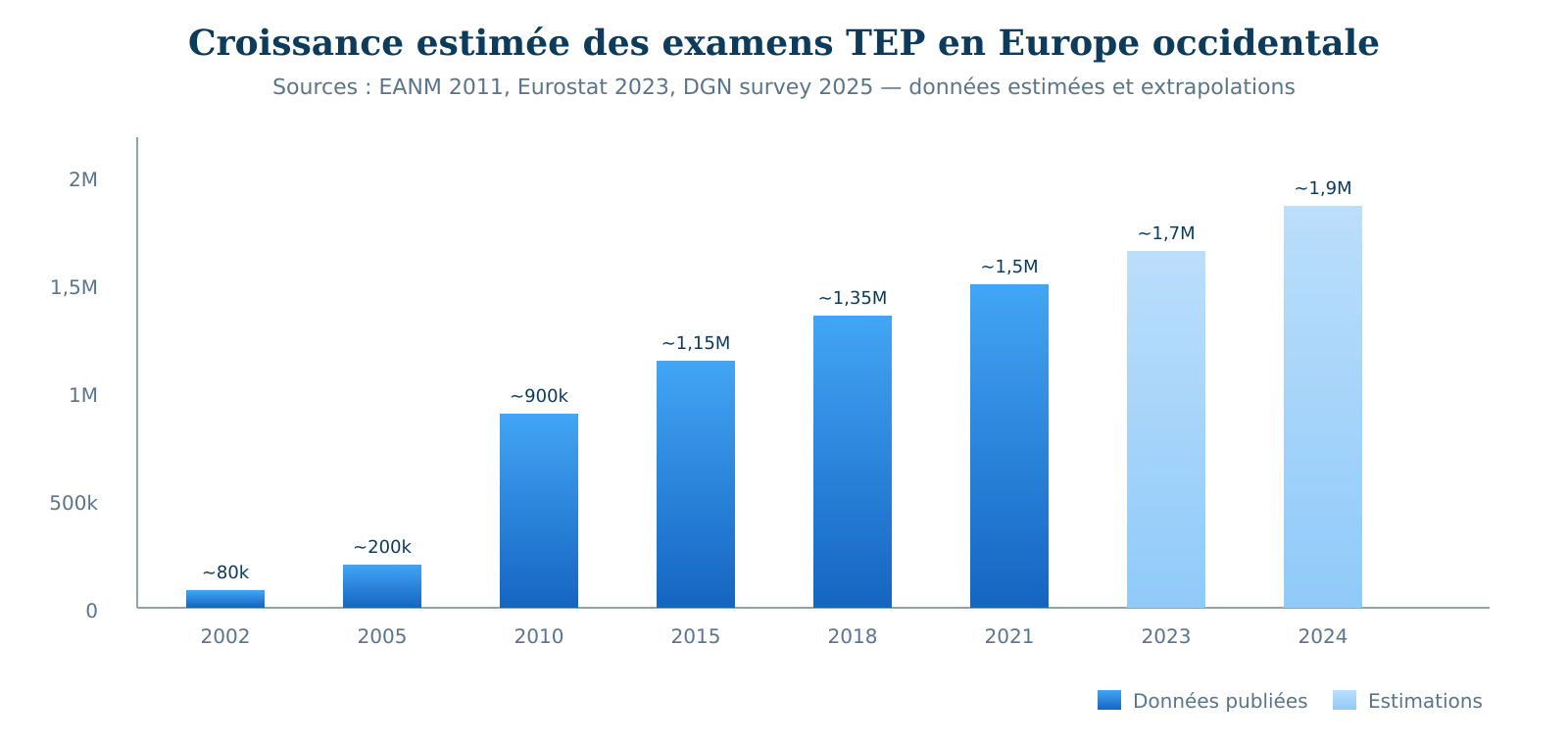
At the turn of the 2000s, PET was still unevenly available in Europe. Equipment needs were already identified, particularly in the United Kingdom, where estimates at the time showed a significant deficit of PET scanners to cover the expected oncological demand.1 The situation changed rapidly with the spread of hybrid PET/CT, which allowed for the fusion of metabolic and anatomical information in a single examination. In Western Europe, examination volumes grew strongly during the 2000s, with an estimated average annual growth of approximately 21% between 2005 and 2010 in certain market analyses presented at the EANM.2
This growth is not the result of technological chance. It results from the convergence of several forces: the rise of hybrid PET/CT systems, the progressive standardization of protocols, the broadening of validated indications, the integration of PET into oncological recommendations, and the arrival of targeted radiotracers beyond ¹⁸F-FDG. ⁶⁸Ga-PSMA, ⁶⁸Ga-DOTATATE, fluorinated PSMA tracers, ¹⁸F-choline and, more recently, FAPI tracers have made it possible to explore tumors with low FDG avidity or specific tumor microenvironments.345
Finally, the emergence of the concept of theranostics — using the same molecular vector to image and then treat — has profoundly modified the role of PET. It is no longer just a staging tool: in several indications, it has become a tool for therapeutic selection and prognostic stratification.6789
This report proposes a structured analysis of twenty years of PET progression in European oncology: usage data, disparities between countries, extension of the tracer spectrum, impact on care pathways, theranostic evolution, and technological perspectives. The objective is not to give in to technological enthusiasm, but to rigorously evaluate what the published data allow us to affirm — and what still remains under evaluation.
In twenty years, PET has moved from the status of a last-resort examination to that of a gold-standard examination in many major oncological indications.
Chronology of an ascent: key figures
The growth of PET in Europe can be divided into three distinct phases, each driven by a different catalyst.
Phase 1 — 2000–2006: the emergence of PET/CT. Before the early 2000s, dedicated PET scanners coexisted with rare cyclotron facilities and still limited radiopharmaceutical logistics. The introduction of hybrid PET/CT systems changed the clinical equation: anatomy and metabolism could finally be superimposed during the same acquisition time, with a major gain for lesion localization, staging, and treatment planning. In the UK, Bedford and Maisey highlighted as early as 2004 the insufficiency of PET supply relative to expected oncological needs.1
Phase 2 — 2006–2016: industrialization. Between 2005 and 2010, data presented at the European Association of Nuclear Medicine congress reported an average growth of approximately 21% per year in PET examination volumes in Western Europe.2 In 2010, 506 centers offered PET or PET/CT activity in this zone, and hybrid PET/CT systems represented 87% of the installed base, compared to 72% in 2006.2 This period corresponds to the industrialization of oncological PET: improved access to ¹⁸F-FDG, multiplication of installations, standardization of protocols, dissemination of EANM and SNMMI recommendations, and gradual recognition of the value of PET/CT in lung cancer, lymphoma, ENT cancer, melanoma, colorectal cancer, and several other indications.10
Phase 3 — 2016–2026: tracer diversification and theranostics. The German study by Holzgreve et al. showed that the national PET volume in Germany reached 154,400 examinations in 2021, 94% of which were PET/CT and 6% PET/MRI.4 German volume had grown by 48% between 2017 and 2021, but remained lower than that observed in several neighboring countries when adjusted for population: 1,857 examinations per million inhabitants in Germany, compared to 10,182 in France, 9,866 in Belgium, and 4,312 in Italy.4
Recent Eurostat data also confirm strong European disparities. In 2023, the European Union had approximately 1,200 PET scanners. France had the largest absolute number of PET scanners, with 233 units recorded, while Denmark had the highest density per inhabitant, with 0.89 PET scanners per 100,000 inhabitants.11 Regarding examinations performed, Denmark, France, and Belgium were among the countries with the highest per capita utilization rates; Denmark reached nearly 3,500 PET examinations per 100,000 inhabitants, which is 2.4 times the French level.11 These figures must be interpreted with caution, as definitions, hospital and extra-hospital scopes, and collection methods vary by country. But the general conclusion is robust: access to PET remains very unequal in Europe.
¹⁸F-FDG: a universal tracer, but not omniscient
¹⁸F-FDG remains the most widely used PET tracer in oncology. Its uptake reflects the glucose hypermetabolism of tumor cells, a phenomenon largely associated with the Warburg effect. This property explains its interest in many aggressive cancers. Validated indications by recommendations include Hodgkin and non-Hodgkin lymphomas, non-small cell lung cancer, melanoma, ENT cancers, colorectal cancer, locally advanced or metastatic breast cancer, ovarian cancer, and many other locations.510
The 2024 publication of joint EANM–SNMMI guidelines on breast cancer consolidated the role of FDG PET/CT in several situations: initial staging of locally advanced forms, therapeutic evaluation, suspected recurrence, and investigation for distant metastases.5 In certain clinical situations, FDG PET/CT can also advantageously replace bone scintigraphy for bone metastatic evaluation, but this substitution must be considered according to the clinical context, tumor stage, and local recommendations.
However, FDG has well-documented limitations. Its accumulation in inflammatory processes can generate false positives, particularly in early post-therapeutic contexts. Certain cancers — well-differentiated hepatocellular carcinoma, low-grade prostate cancer, certain well-differentiated neuroendocrine tumors, small bowel carcinoids — may be poorly or not avid for FDG. It is precisely to fill these gaps that multi-tracer PET has developed.
The explosion of targeted tracers: PSMA, DOTATATE, FAPI and beyond
The 2014–2024 decade was marked by the introduction into clinical practice of highly specific tracers, transforming the diagnostic strategy for several cancers.
PSMA: the paradigm shift in prostate cancer. ⁶⁸Ga-PSMA and its fluorinated equivalents, such as ¹⁸F-DCFPyL or ¹⁸F-PSMA-1007, target the prostate-specific membrane antigen, overexpressed in many prostate cancers. ⁶⁸Ga-PSMA-11 was approved by the FDA on December 1, 2020, as the first PSMA-targeted PET agent in prostate cancer.12 In Europe, the Locametz® kit, after radiolabeling with gallium-68, is indicated for the detection by PET of PSMA-positive lesions in adults with prostate cancer in several clinical contexts.13 In the 2021 German study, prostate cancer already accounted for 16% of all PET indications, behind lung indications but ahead of neurology and inflammatory pathologies.4
DOTATATE: the reference in neuroendocrine tumors. ⁶⁸Ga-DOTATATE and its analogs, notably DOTATOC and DOTANOC, target somatostatin receptors, overexpressed in many well-differentiated neuroendocrine tumors. PET with somatostatin analogs has largely supplanted Octréoscan® scintigraphy in this indication, thanks to better resolution, better sensitivity, and better lesion characterization. This imaging also plays a central role in selecting patients for ¹⁷⁷Lu-DOTATATE. The NETTER-1 trial demonstrated the benefit of ¹⁷⁷Lu-DOTATATE in progressive intestinal neuroendocrine tumors expressing somatostatin receptors.8 Lutathera® is authorized in Europe for well-differentiated, progressive, unresectable or metastatic gastroenteropancreatic neuroendocrine tumors expressing somatostatin receptors.9
FAPI: a promising frontier but still under evaluation. Tracers targeting fibroblast activation protein — FAPI — represent one of the most recent frontiers of oncological molecular imaging. Their potential interest is based on targeting cancer-associated fibroblasts, present in the tumor stroma of many solid tumors. Clinical data have shown the interest of FAPI-PET in several tumor entities, with a single-center database of 324 patients and 21 tumor types published in the Journal of Nuclear Medicine.3 Therapeutic approaches using ⁹⁰Y-FAPI-46 have also been explored in advanced cancers, notably sarcomas and pancreatic cancer, with encouraging preliminary results but still insufficient to define established clinical efficacy.14 Caution is therefore required: FAPI tracers do not yet have broad European authorization in common oncological indications, and their use remains mainly in the field of clinical research, early trials, or institutional protocols.
⚠ Attention. The extension of indications for new tracers must be interpreted with caution. Long-term survival data, standardized response criteria, cost-effectiveness analyses, and reimbursement recommendations are still maturing for many of them. Multi-tracer PET increases the information available, but also decision complexity and the risk of overinterpretation.
Impact on care pathways: what the data say
One of the strongest arguments in favor of extending PET is its ability to modify the therapeutic plan. In aggressive lymphomas, post-chemotherapy PET/CT influences the decision for consolidation irradiation, notably through the Deauville score. In non-small cell lung cancer, it can change the TNM stage, avoid certain unnecessary thoracotomies, or conversely, identify patients for whom an initially planned surgical strategy is no longer suitable. In locally advanced breast cancer, the metabolic response under neoadjuvant chemotherapy can provide complementary prognostic information.
The direct impact on survival is more difficult to establish, due to biases inherent in any staging tool: stage migration, lead-time bias, concomitant improvement in treatments, and evolution of care pathways. Ecological data nevertheless remain suggestive. In the international study by Lynch et al., the improvement in the 1-year survival rate for lung cancer in Denmark — from 27.5% over the 1995–1999 period to 46.2% over 2010–2014 — is discussed in the context of better cancer pathway structuring and increased access to PET/CT.15 This association does not prove direct causality, but it reinforces the clinical coherence of organized access to modern imaging.
Theranostics constitutes the most structuring evolution. PSMA-PET allows for the identification of patients with metastatic castration-resistant prostate cancer expressing PSMA, who are likely to benefit from treatment with ¹⁷⁷Lu-PSMA-617. The VISION trial showed that adding ¹⁷⁷Lu-PSMA-617 to the standard of care prolonged overall survival and radiographic progression-free survival in pretreated PSMA-positive mCRPC.7 The EMA authorizes Pluvicto® in metastatic, progressive, castration-resistant, PSMA-positive prostate cancer, following appropriate systemic treatments.6 Similarly, somatostatin receptor imaging selects neuroendocrine tumor patients for ¹⁷⁷Lu-DOTATATE.89
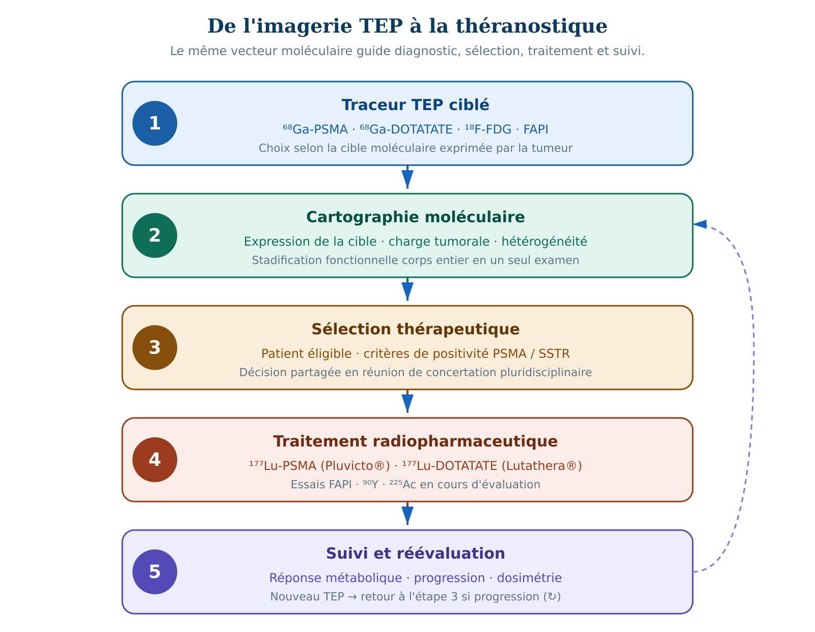
📌 Key takeaway. PET is no longer just a staging tool. In several indications, it has become a therapeutic selection tool, particularly for targeted radiopharmaceutical treatments.
European disparities and issues of access equity
Eurostat 2023 data reveal considerable inequalities in access to PET equipment and examinations. The European Union had approximately 1,200 PET scanners, with a high concentration in France, Germany, and Italy in absolute numbers.11 However, density per inhabitant and the number of examinations performed vary greatly between countries. Denmark stands out with a very high utilization rate, with nearly 3,500 PET examinations per 100,000 inhabitants in 2023. France and Belgium also figure among the European countries with the highest utilization rates, while certain Central, Eastern, or Northern European countries show much lower rates.11
This inequality is not just a problem of technical capacity. It can represent a difference in care quality in pathologies where PET/CT is part of the international standard. Access to PET/CT conditions the diagnosis, staging, therapeutic decision, and, in theranostic indications, access to certain radiopharmaceutical treatments.
The EANM Position Paper on Nuclear Medicine identifies several structural obstacles: shortage of trained nuclear medicine physicians, heterogeneity of reimbursement policies, difficulties in radiotracer supply, infrastructure limits, regulatory burden, and lack of strategic recognition of nuclear medicine in some health systems.16 The issue of reimbursement is central. In France and Belgium, relatively high utilization levels reflect more favorable access policies. Conversely, Germany, despite its economic development level, shows a lower utilization rate in 2021 comparative data, partly due to historical reimbursement constraints and healthcare system organization.4
Emerging technologies: Total-body PET and artificial intelligence
Total-body PET and long axial field-of-view systems. Total-body PET or long axial field-of-view PET systems appeared in the late 2010s with early systems like EXPLORER/uEXPLORER.1718 Their principle is based on much longer axial coverage than conventional PET, allowing more photons to be captured and strongly increasing the system's global sensitivity. Initial human studies with EXPLORER showed the feasibility of simultaneous total-body acquisition.17 Technical evaluations report, depending on systems and comparisons, sensitivity multiplied by approximately 15 to 60 times compared to conventional PET.18
In practice, this increased sensitivity opens several perspectives: reduction of injected activity, faster acquisition, dynamic total-body imaging, better kinetic quantification, and exploration of fragile or pediatric populations with potentially reduced dosimetry. These systems however remain expensive, few in number, and still mainly concentrated in academic or research centers. Their real clinical impact on routine management remains to be established by multicenter studies.
Artificial intelligence. Artificial intelligence is gradually integrating into the PET image production and interpretation chain: accelerated reconstruction, denoising, motion correction, automatic segmentation of tumor volumes, measurement of MTV and TLG, computer-aided lesion detection, and predictive modeling of therapeutic response. These tools offer significant potential, but their clinical deployment requires rigorous external validation, evaluation of inter-center robustness, management of training biases, and compliance with European requirements applicable to software medical devices. Automatic segmentation can improve reproducibility, but it must not be confused with an autonomous clinical decision.
Perspectives 2026–2035: what the data allow us to anticipate
The European PET scanner market is estimated at approximately $241 million in 2024, with a projection to $321 million in 2033 according to certain market analyses, i.e., at a moderate compound annual growth of approximately 3.21%.19 This equipment market growth however does not summarize the actual clinical dynamic, which will depend more on indication extension, radiotracer access, team availability, reimbursement, and theranostic integration.
Several evolutions should be monitored closely. FAPI-PET, if prospective trials confirm its diagnostic and theranostic interest, could open new patient pools, notably in certain cancers with low FDG avidity or characterized by an abundant tumor stroma. Tracers targeting estrogen receptors, androgen receptors, or tumor hypoxia could refine therapeutic selection in certain mammary, prostate, or radio-resistant tumors. Carbon-11 PET, notably ¹¹C-choline or ¹¹C-acetate, will likely remain limited to centers with an on-site cyclotron, due to the very short half-life of carbon-11.
The growth of PET is therefore not just a quantitative increase in acts. It reflects a qualitative transformation of the role of nuclear medicine: from complementary functional imaging towards a central tool of precision oncology.
📌 Key takeaway. The PET of tomorrow will be defined less by a single universal tracer than by a personalized molecular strategy: choosing the right tracer, for the right patient, at the right therapeutic moment.
In twenty years, PET has moved from the periphery to a central position in many oncological pathways. The data are convergent: rapid growth in examination volumes during the 2000s, dissemination of hybrid PET/CT systems, extension of validated indications, multiplication of targeted radiotracers, and gradual integration of molecular imaging into theranostic strategies. However, challenges remain considerable. Access inequalities within Europe are deep and documented. Regulation of new radiotracers, pharmacovigilance, radiopharmaceutical traceability, and requirements applicable to interpretation software impose increasing operational constraints. The integration of AI and high-sensitivity systems raises questions of clinical validation, reproducibility, and medical-legal responsibility. Finally, the increasing complexity of diagnostic options requires nuclear medicine teams to have decision-making expertise and permanent scientific monitoring. PET is therefore not a technique in passive growth. It is in active transformation, driven by research, pulled by theranostics, and facing issues of equity, regulation, and sustainability that the European nuclear medicine community must continue to address collectively.
The PET of 2026 is no longer the diagnostic tool of 2006: it has become an entry point into a molecular imaging and theranostic ecosystem of which we are only beginning to explore the first layers.
🔑 Key points to remember • Growth 2005–2010 — PET progressed rapidly in Western Europe during the 2000s, with an estimated average annual growth of approximately 21% between 2005 and 2010 in certain analyses presented at the EANM. • Intra-European disparity — Eurostat 2023 data show strong disparities: Denmark, France, and Belgium are among the countries with the highest utilization per inhabitant, while several countries remain significantly below. • FDG dominance — ¹⁸F-FDG remains the central tracer for PET oncological imaging, but it is neither universal nor sufficient for all tumors. • PSMA on the rise — PSMA has transformed prostate cancer imaging, notably in biological recurrence, staging of high-risk forms, and patient selection for radioligand treatments. • DOTATATE and NET — Somatostatin receptor imaging has become essential in well-differentiated neuroendocrine tumors and in selecting patients for ¹⁷⁷Lu-DOTATATE. • Theranostics — PET now plays a direct role in access to certain radiopharmaceutical therapies, notably ¹⁷⁷Lu-PSMA-617 and ¹⁷⁷Lu-DOTATATE. • Total-body PET — Long axial field-of-view systems offer a much higher sensitivity than conventional systems, but their dissemination remains limited and their clinical impact must still be validated on a large scale. • FAPI under evaluation — FAPI tracers constitute a promising frontier, but their clinical role is still mostly under evaluation. • AI and quantification — Artificial intelligence could improve reconstruction, segmentation, and quantification, but still requires rigorous external validation before broad routine deployment.
📑 References • Bedford M, Maisey MN. Requirements for clinical PET: comparisons within Europe. Eur J Nucl Med Mol Imaging. 2004;31:208–221. doi:10.1007/s00259-003-1351-6 • Stevens A. EANM: PET and PET/CT scans grow by 21% a year. AuntMinnieEurope. Oct 21, 2011. Data from a presentation at the EANM Annual Congress, Birmingham, 2011. • Hirmas N, Hamacher R, Sraieb M, et al. Fibroblast-Activation Protein PET and Histopathology in a Single-Center Database of 324 Patients and 21 Tumor Entities. J Nucl Med. 2023;64(5):711–716. doi:10.2967/jnumed.122.264689 • Holzgreve A, Hellwig D, Barthel H, et al. PET imaging utilization and trends in Germany: a comprehensive survey. Eur J Nucl Med Mol Imaging. 2025. doi:10.1007/s00259-025-07323-x • Vaz SC, Pilkington Woll JP, Cardoso F, Groheux D, Cook GJR, Ulaner GA, et al. Joint EANM–SNMMI guideline on the role of 2-[¹⁸F]FDG PET/CT in no special type breast cancer. Eur J Nucl Med Mol Imaging. 2024;51:2706–2732. doi:10.1007/s00259-024-06696-9 • European Medicines Agency. Pluvicto: EPAR — Product information. EMA; initial European authorization 2022, subsequent updates. • Sartor O, de Bono J, Chi KN, et al. Lutetium-177–PSMA-617 for Metastatic Castration-Resistant Prostate Cancer. N Engl J Med. 2021;385:1091–1103. doi:10.1056/NEJMoa2107322 • Strosberg J, El-Haddad G, Wolin E, et al. Phase 3 Trial of ¹⁷⁷Lu-Dotatate for Midgut Neuroendocrine Tumors. N Engl J Med. 2017;376:125–135. doi:10.1056/NEJMoa1607427 • European Medicines Agency. Lutathera: EPAR — Product information. EMA; initial European authorization 2017, subsequent updates. • Boellaard R, Delgado-Bolton R, Oyen WJG, et al. FDG PET/CT: EANM procedure guidelines for tumour imaging: version 2.0. Eur J Nucl Med Mol Imaging. 2015;42:328–354. doi:10.1007/s00259-014-2961-x • Eurostat. Healthcare resource statistics — technical resources and medical technology. 2023 data; datasets hlth_rs_medim and hlth_co_exam. Eurostat Statistics Explained. • U.S. Food and Drug Administration. FDA approves first PSMA-targeted PET imaging drug for men with prostate cancer. FDA; Dec 1, 2020. • European Medicines Agency. Locametz: EPAR — Product information. EMA; initial European authorization 2022, subsequent updates. • Fendler WP, Pabst KM, Kessler L, et al. Safety and Efficacy of ⁹⁰Y-FAPI-46 Radioligand Therapy in Patients with Advanced Sarcoma and Other Cancer Entities. Clin Cancer Res. 2022;28:4346–4353. doi:10.1158/1078-0432.CCR-22-1432 • Lynch C, Reguilon I, Langer DL, et al. A comparative analysis: international variation in PET-CT service provision in oncology — an International Cancer Benchmarking Partnership study. Int J Qual Health Care. 2021;33(1):mzaa166. doi:10.1093/intqhc/mzaa166 • European Association of Nuclear Medicine. Nuclear Medicine — What it is. Where it goes. What it needs. EANM position paper / overarching narrative. 2022–2024. • Badawi RD, Shi H, Hu P, et al. First Human Imaging Studies with the EXPLORER Total-Body PET Scanner. J Nucl Med. 2019;60(3):299–303. doi:10.2967/jnumed.119.226498 • Spencer BA, Berg E, Schmall JP, et al. Performance Evaluation of the uEXPLORER Total-Body PET/CT Scanner Based on NEMA NU 2-2018 with Additional Tests to Characterize PET Scanners with a Long Axial Field of View. J Nucl Med. 2021;62(6):861–870. doi:10.2967/jnumed.120.250597 • Market Data Forecast. Europe PET Scanners Market Size, Share & Growth Forecast to 2033. Market report, 2026 edition.