Artificial intelligence in nuclear medicine
Month : 2026-04
Diagnostic support, image enhancement and reporting assistance. A concrete and measured overview of current AI applications in our specialty, in light of 2024–2026 publications.
Nuclear medicine is already a highly technological field. It relies on imaging, quantification, reconstruction software, clinical data and, increasingly, automation. In this context, artificial intelligence does not arrive as a total disruption, but rather as a new layer of assistance, capable of accelerating certain tasks, improving image quality and supporting interpretation.
Since 2024, the specialist literature has shown a clear acceleration in work on AI applied to PET, SPECT, artefact correction, segmentation, quantification and assisted report writing. Several major reviews — in Radiological Physics and Technology1, the Journal of Nuclear Medicine2 and Annals of Nuclear Medicine3 — now provide a coherent overview of real contributions and persistent limitations.
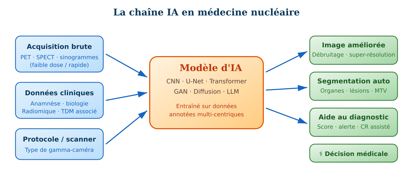
AI can help nuclear medicine be faster, more reproducible and sometimes more precise. But it is neither magical nor autonomous.
01 · Diagnostic support
In nuclear medicine, AI is particularly well-suited to tasks requiring the detection of subtle patterns, comparison of intensities, measurement of volumes or integration of large amounts of information. It is therefore heavily studied in oncology, neurology and nuclear cardiology.
In practice, AI can help detect a difficult-to-see lesion more effectively, estimate tumour burden faster, automatically segment structures or standardise certain quantitative measurements. In neurodegenerative diseases, it helps analyse uptake patterns in amyloid or tau PET.
A notable example published in early 2026 by Zhang et al. in the European Journal of Nuclear Medicine and Molecular Imaging illustrates the value of a 3D multi-modal PET/CT model trained on 474 patients: for T-stage prediction of colorectal cancer, the model combining PET+CT+clinical data achieves an AUC of 0.869 in internal validation, outperforming single-modality models4.
Key point. The algorithm does not make the diagnosis in place of the physician. It acts as a digital second reader: it draws attention, pre-segments, quantifies, proposes a classification or score. The final diagnosis remains clinical and human.
02 · Cleaner and more usable images
One of the areas where AI is most impressive today is image enhancement. Deep learning models are capable of improving noisy PET or SPECT images, restoring low-count acquisitions and sometimes making usable examinations acquired faster or at lower dose.
For the patient, the potential benefit is very concrete. If a good-quality image can be obtained with less noise despite fewer counts, this opens the door to a reduction in injected dose or a shorter acquisition time.
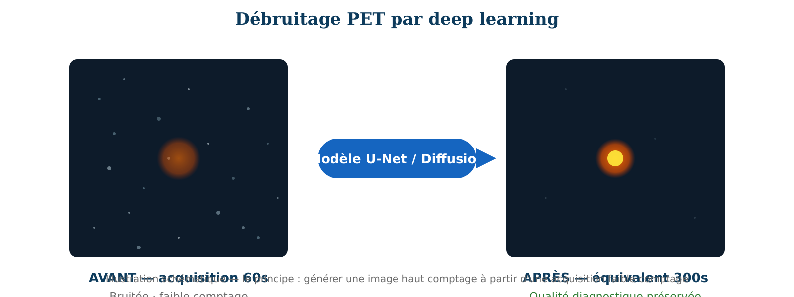
This principle is now validated in large-scale multicentre studies. Maes et al. published in 2025 in EJNMMI a blinded external evaluation of NUCLARITY software on 65 examinations acquired in three European hospitals with four different radiotracers; six independent nuclear medicine physicians judged the diagnostic quality of 50%-reduced-count images as equivalent to standard images6.
However, caution is warranted. A visually more beautiful image is not always synonymous with perfect biological fidelity. A study of 110 patients showed that tumour response classifications differed in 10% of patients when using SubtlePET-processed FDG scans versus conventional reconstruction3. Practical conclusion: never mix denoised and non-denoised images in longitudinal follow-up.
03 · Correcting what the eye does not always see
AI is not only used to clean up the final image. It also intervenes earlier in the technical chain: respiratory and cardiac motion correction, attenuation correction (including without associated CT via synthetic atlases for PET/MRI), automatic PET-CT co-registration, synthesis of images useful for reconstruction, and scatter correction.
04 · Automatic segmentation: the silent revolution
Segmentation — precisely delineating the contours of an organ or lesion in a 3D image — is a crucial but extraordinarily time-consuming task. Manually delineating all tumour lesions in a whole-body lymphoma scan can take an experienced nuclear medicine physician more than an hour. AI is radically transforming this step.
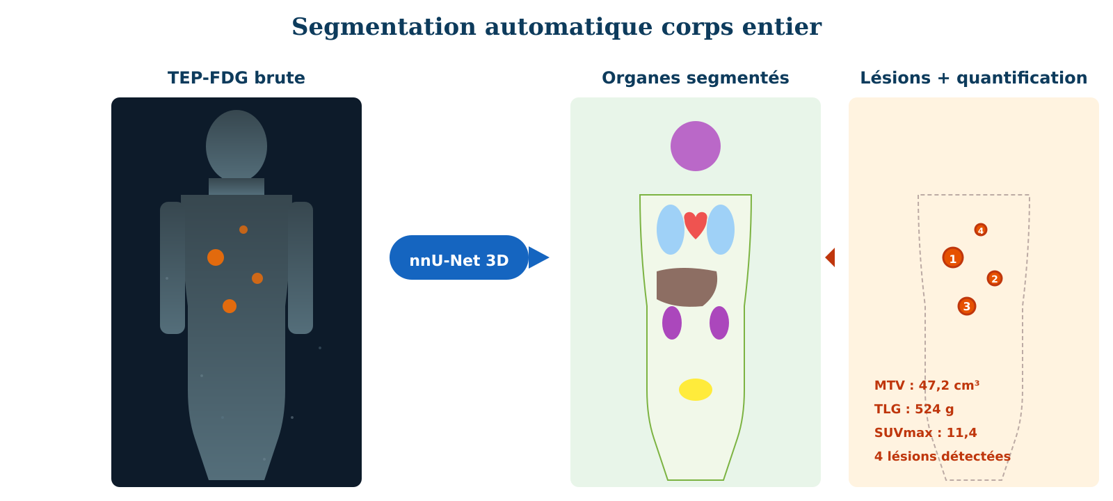
Generalist models such as TotalSegmentator (segments 117 anatomical structures on CT)11, MOOSE (dedicated to whole-body PET/CT) and especially recent foundation models such as SegAnyPET12 or VISTA3D13 are now available as open-source or semi-commercial. They have significantly superior generalisation capability compared to older organ-specific models.
05 · Assistance in report writing
Large language models (LLMs) are increasingly studied in nuclear medicine to assist writing, structure reports, summarise findings, extract key data (SUVmax, location, size) or transform free text into more standardised information.
Warning. This is the area where the risk of credible error is most concerning. A language model can produce a very well-written but factually false sentence. These tools must remain editorial assistants under strict medical supervision, with systematic word-by-word proofreading by the signing physician. Riedemann et al. (2024) detail the safeguards necessary for their deployment in medicine14.
06 · Better linking image to clinical data
AI becomes particularly interesting when it fuses multiple sources: PET or SPECT image, associated CT, clinical data, sometimes molecular biology. It can help link an image to a tumour context, a risk of relapse, an expected response to treatment. This is the principle of multi-modal deep radiomics.
07 · Current limitations and future challenges
• Lack of well-annotated multicentre data. Most models are trained on single-centre cohorts and lose performance when applied to images acquired on different machines with different protocols. Domain shift remains the Achilles heel of deep learning in medical imaging. • Variations between machines and protocols. A model trained on a Siemens Biograph Vision scanner often performs less well on an older GE Discovery PET. • Regulatory constraints and GDPR. Placing an AI medical device on the market requires CE marking (MDR 2017/745 regulation), often in class IIa or IIb. • Reproducibility and explainability. A "black box" algorithm that simply says "abnormal" without justifying its decision is difficult to accept in a medical report. • Training and acculturation of teams. Sustainable adoption of an AI tool in clinical practice requires initial and ongoing training of physicians and technicians.
Conclusion AI in nuclear medicine is no longer a futuristic idea. It is already present, especially in image enhancement, PET and SPECT denoising, automatic segmentation, quantification and reporting assistance. Publications 2024-2026 mark a clear transition: from proof of concept to external multicentre clinical validation. AI does not replace the nuclear medicine specialist, but it can save them time, improve the technical quality of certain examinations, reduce dose or acquisition time, and make analysis more homogeneous between readers. The challenge for our specialty over the next five years is less technical than cultural and organisational: knowing how to choose tools that bring real clinical value, validate them locally and train teams. AI is probably becoming a very good co-pilot for nuclear medicine. And like any good co-pilot, its value comes not from its ability to decide alone, but from its ability to intelligently assist the professional who retains ultimate responsibility.
🔑 Key points • Diagnostic support — AI detects subtle patterns, pre-segments lesions, quantifies tumour burden and standardises measurements; it acts as a digital second reader, not a decision-maker. • Image enhancement — deep learning denoises PET and SPECT acquisitions, opening the way to dose reduction or shorter acquisition time; validated in multicentre multi-tracer studies (NUCLARITY, SubtlePET, AiCE, etc.). • Pre-reconstruction correction — AI corrects motion, attenuation and PET-CT co-registration, for more reliable dosimetry and therapeutic monitoring, particularly in RLT. • Automatic segmentation — foundation models (TotalSegmentator, MOOSE, SegAnyPET, VISTA3D) now segment 100+ structures in under a minute, finally making MTV and TLG practicable in routine. • Reporting assistance — LLMs structure reports and extract key data, but must remain under strict medical supervision (risk of plausible factual error). • Multimodal fusion — AI links molecular image, CT, clinical data and biology to anticipate relapse risk and therapeutic response. • Current limitations — few tools transition from prototype to broad clinical use: inter-machine domain shift, lack of annotated data, MDR/FDA and GDPR constraints. • Final responsibility — clinical diagnosis and therapeutic decision-making remain human; AI is a co-pilot, not a pilot.
• Hashimoto F, Onishi Y, Ote K, Tashima H, Reader AJ, Yamaya T. Deep learning-based PET image denoising and reconstruction: a review. Radiol Phys Technol. 2024;17(1):24-46. doi:10.1007/s12194-024-00780-3 • Balaji V, Song TA, Malekzadeh M, Heidari P, Dutta J. Artificial Intelligence for PET and SPECT Image Enhancement. J Nucl Med. 2024;65(1):4-12. doi:10.2967/jnumed.122.265000 • Miwa K, et al. Innovations in clinical PET image reconstruction: advances in Bayesian penalized likelihood algorithm and deep learning. Ann Nucl Med. 2025. doi:10.1007/s12149-025-02088-7 • Zhang M, Li Y, Zheng C, et al. Fully automated 3D multi-modal deep learning model for preoperative T-stage prediction of colorectal cancer using 18F-FDG PET/CT. Eur J Nucl Med Mol Imaging. 2026;53:910-920. doi:10.1007/s00259-025-07450-5 • Zhang MX, Liu PF, Zhang MD, et al. Deep learning in nuclear medicine: from imaging to therapy. Ann Nucl Med. 2025;39(5):424-440. • Maes J, Carron C, DeKeyser S, et al. Low-count whole-body PET denoising with deep learning in a multicenter, multi-tracer and externally validated study. Eur J Nucl Med Mol Imaging. 2025. doi:10.1007/s00259-025-07672-7 • Silva LC, Constantino C, Teixeira R, et al. Assessing the quantitative feasibility of deep-learning-based denoising of faster-than-1-minute whole-body [18F]FDG PET acquisitions. J Nucl Med. 2025;66(suppl 1):251342. • PET Image Denoising with Rectified Flow. J Nucl Med. 2025;66(suppl 1):251475. • Xue S, Liu F, Wang H, et al. A deep learning method for the recovery of standard-dose imaging quality from ultra-low-dose PET on wavelet domain. Eur J Nucl Med Mol Imaging. 2025;52:1901-1911. doi:10.1007/s00259-024-06994-2 • Raymond C, Zhang D, Cabello J, et al. SMART-PET: a Self-SiMilARiTy-aware generative adversarial framework for reconstructing low-count [18F]-FDG-PET brain imaging. Front Nucl Med. 2024;4:1469490. doi:10.3389/fnume.2024.1469490 • Wasserthal J, Breit HC, Meyer MT, et al. TotalSegmentator: Robust Segmentation of 104 Anatomic Structures in CT Images. Radiol Artif Intell. 2023;5(5):e230024. doi:10.1148/ryai.230024 • Zhang Y, Xue L, Zhang W, et al. SegAnyPET: universal promptable segmentation from positron emission tomography images. Proc IEEE/CVF ICCV. 2025:21107-21116. • He Y, et al. VISTA3D: A Unified Segmentation Foundation Model For 3D Medical Imaging. Proc IEEE/CVF CVPR. 2025. • Riedemann L, Labonne M, Gilbert S. The path forward for large language models in medicine is open. npj Digit Med. 2024;7:339. doi:10.1038/s41746-024-01344-w • Kersting D, Borys K, Küper A, et al. Staging of prostate cancer with ultra-fast PSMA-PET scans enhanced by AI. Eur J Nucl Med Mol Imaging. 2025;52(5):1658-1670.